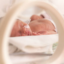
An infant with congenital heart disease was deteriorating at Lucile Packard Children's Hospital Stanford despite multiple open-heart surgeries. His heart was enlarged and not pumping properly, but traditional imaging couldn’t explain why.
A high-resolution MRI technique built on AI modeling revealed a subtle abnormality the other imaging couldn’t see. The surgeons quickly corrected it, and the child recovered.
“This diagnosis would have been nearly impossible without these new techniques,” said Michael Ma, MD, the baby’s surgeon and division chief of pediatric cardiac surgery.
The same technology is reshaping pediatric imaging in less dramatic but more routine ways every day. It reduces scan times from an hour to minutes, decreases reliance on anesthesia, shortens wait times, and, in cases like this one, reveals detail that older methods miss.
Solving a pediatric problem
Conventional MRI presents unique challenges in pediatrics.
Motion introduces artifacts that can make scans difficult to interpret, particularly during lengthy exams. As a result, many kids require anesthesia, which adds cost, complexity, scheduling constraints, and potential risks.
The technology carries its own limits. Most MRI systems and hardware are designed for adults, including the receiver coils that capture MRI signals. Those adult-centered designs sacrifice image quality and efficiency on smaller anatomical structures.
AI offered a potential solution. Instead of collecting every piece of MRI data, could an algorithm learn how to reconstruct high-quality images from a fraction of the information?
The answer turned out to be yes.
Training AI to do more with less
Shreyas Vasanawala, MD, PhD, Stanford Medicine Children’s Health’s radiologist-in-chief and chief of the division of pediatric radiology at the Stanford University School of Medicine, developed deep-learning approaches that train algorithms using completed MRI studies. Once a model learns what a fully reconstructed image should look like, it can build one from a very small subset of the data.
“If you collect only 10% of the data, you image 10 times as fast,” Vasanawala said.
Congenital heart exams that once took more than an hour now finish in about 10 minutes. Some orthopedic and follow-up brain studies take as little as five.
For many children, that means avoiding anesthesia altogether.
“They won’t give you an hour of cooperation, but they may give you five minutes,” Vasanawala said.
Even when anesthesia is still needed, shorter exams allow for lighter and briefer sedation.
Faster scans also mean more scans, which raises throughput for the hospital and improves access for patients.
“Historically, many of our patients are driving two or more hours to reach us. They see an orthopedic surgeon, and then they’re told to go home and wait for a call to get scheduled to come right back here for an MRI,” Vasanawala said. “The parents now have to take another day off work, the child another day out of school, just to get this scan. So it’s a huge patient satisfier if they can simply get a walk-in MRI appointment.”
Enhanced imaging
Hospitals can use the technology to shorten scans, or they can trade that speed for higher resolution and evaluate extremely small structures.
Infants with anorectal malformations, for example, may have an abnormal connection between the urinary and gastrointestinal systems smaller than a millimeter across. Higher-resolution imaging can see it.
In musculoskeletal imaging, Stanford’s AI-enhanced knee MRI protocols produce slices 600 microns thick, compared with the 2.5- to 3-millimeter slices in conventional studies. The additional detail allows radiologists to evaluate structures like the anterior cruciate ligament with far greater precision, including individual fibers.
“When we started using this as radiologists, we had to go back and learn some more detailed anatomy, because we were seeing things and asking, ‘What is this structure?’” Vasanawala said.
Improving workflow
Vasanawala is also aiming AI at the mundane, manual work of operations.
Working with Drs. Sergios Gatidis, Ali Syed, Liliana Ma, and data systems engineer Amol Sinha, the team has incorporated large language models that help staff navigate more than 1,200 pages of departmental protocols, policies, workflows, and scheduling guidance. Instead of searching through multiple documents, technologists, schedulers, and clinicians ask questions in plain language and get answers linked to the source.
The department also uses AI to flag discrepancies between preliminary and final radiology reports. That improves communication with referring providers and creates teaching moments for residents and fellows.
Vasanawala sees similar opportunities in exam scheduling, protocol selection, and resource management.
“When you order a brain MRI, we’ve got 30 different protocols,” he said.
AI could help automate many of those decisions while improving consistency and efficiency.
Where to begin
Vasanawala’s team also helped port these algorithms to GE MRI scanners, which have now been used at 5,000 sites across 160 countries.
He recommends approaching AI as hospitals would any other clinical technology: Understand how it works, monitor quality carefully, and implement it gradually.
“You can misuse any technology,” he said. “AI is no exception.”
Successful deployment requires data consistency checks, quality assurance, and continuous oversight.
For Vasanawala, these advances are only the beginning.
“It’s a fantastic time,” he said. “The potential for transforming patient care and outcomes is almost limitless.”